


General Information About Esophageal Cancer
- Esophageal cancer is a disease in which malignant (cancer) cells form in the tissues of the esophagus.
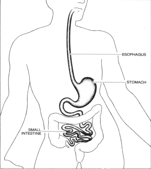
- The esophagus is the hollow, muscular tube that moves food and liquid from the throat to the stomach. The wall of the esophagus is made up of several layers of tissue, including mucous membrane, muscle, and connective tissue. Esophageal cancer starts on the inside lining of the esophagus and spreads outward through the other layers as it grows.
- Smoking, heavy alcohol use, and Barrett esophagus can increase the risk of esophageal cancer.
- Signs and symptoms of esophageal cancer are weight loss and painful or difficult swallowing
Types of Esophageal Cancer
The two most common forms of esophageal cancer are named for the type of cells that become malignant (cancerous):
Squamous cell carcinoma: Cancer that forms in the thin, flat cells lining the inside of the esophagus. This cancer is most often found in the upper and middle part of the esophagus, but can occur anywhere along the esophagus.
Adenocarcinoma: Cancer that begins in glandular cells. Glandular cells in the lining of the esophagus produce and release fluids such as mucus. Adenocarcinomas usually form in the lower part of the esophagus, near the stomach.
Risk factors for esophageal cancer
- Tobacco use.
- Heavy alcohol use.
- Barrett esophagus: A condition in which the cells lining the lower part of the esophagus have changed or been replaced with abnormal cells that could lead to cancer of the esophagus. Gastric reflux (heartburn) is the most common cause of Barrett esophagus.
- Older age.
Signs and symptoms of esophageal cancer
- Painful or difficult swallowing.
- Weight loss.
- Pain behind the breastbone.
- Hoarseness and cough.
- Indigestion and heartburn.
- A lump under the skin.
Tests used to diagnose esophageal cancer
- Physical exam and health history
- Esophagoscopy: A procedure to look inside the esophagus to check for abnormal areas. An esophagoscope is inserted through the mouth or nose and down the throat into the esophagus. An esophagoscope is a thin, tube-like instrument with a light and a lens for viewing. It may also have a tool to remove tissue samples, which are checked under a microscope for signs of cancer. When the esophagus and stomach are looked at, it is called an upper endoscopy.
- MRI (magnetic resonance imaging): A procedure that uses a magnet, radio waves, and a computer to make a series of detailed pictures of areas inside the body.
- CT scan (CAT scan): A procedure that makes a series of detailed pictures of areas inside the body, taken from different angles. The pictures are made by a computer linked to an x-ray machine.
- PET scan (positron emission tomography scan): A procedure to find malignant tumor cells in the body. A small amount of radioactive glucose (sugar) is injected into a vein. Malignant tumor cells show up brighter in the picture because they are more active and take up more glucose than normal cells do. A PET scan and CT scan may be done at the same time. This is called a PET-CT.
- Abdominal ultrasound: An ultrasound exam used to make pictures of the inside of the abdomen.
- Endoscopic ultrasound (EUS): A procedure in which an endoscope is inserted into the body, usually through the mouth. An endoscope is a thin, tube-like instrument with a light and a lens for viewing. A probe at the end of the endoscope is used to bounce high-energy sound waves (ultrasound) off internal tissues or organs and make paictures.
- Biopsy
Stages of Esophageal Cancer
Tests and procedures to stage esophageal cancer are usually done at the same time as diagnosis.
There are three ways that cancer spreads: direct spread to neighboring tissue, through blood stream and through lymph channels.
The following stages are used for Esophageal cancer:
- Stage 0 (High-grade Dysplasia)
- Stage I
- Stage II
- Stage III
- Stage IV
Grade of Esophageal Cancer
The grade of the tumor describes how abnormal the cancer cells look under a microscope and how quickly the tumor is likely to grow and spread.
- Grades 1 to 3 are used to describe esophageal cancer.
- In grade 1, the cancer cells look more like normal cells under a microscope and grow and spread more slowly than grade 2 and 3 cancer cells.
- In grade 2, the cancer cells look more abnormal under a microscope and grow and spread more quickly than grade 1 cancer cells.
- In grade 3, the cancer cells look even ore abnormal under a microscope and grow and spread more quickly than grade 1 and 2 cancer cells.
Additional information can be found at: https://www.cancer.gov/types/esophageal/patient/esophageal-treatment-pdq


